Overview of the technique
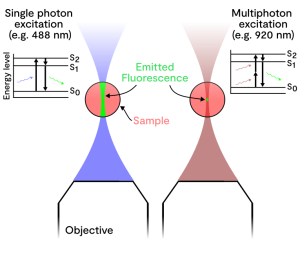
Laser scanning multiphoton microscopy offers advantages over wide-field and confocal fluorescence microscopy when using thick samples, such as tissues, and high fluorophore labelling densities. The excitation light is usually two or three times longer in wavelength than widefield or confocal, two-photon or three-photon excitation respectively, with less energy and reduced scattering in the sample for deeper imaging. The technique generates fluorescence excitation from only a very small focal volume at the focal plane, reducing any out of focus light and increasing signal-to-noise. This is due to the unlikely event of multiphoton excitation, first proposed by Maria Göppert-Mayer, arising from simultaneous absorption of two photons in a single event within the order of one attosecond, which only happens at the focal volume with high photon density.
Multiphoton imaging therefore provides optical sectioning capabilities to depths of greater than 1mm at diffraction-limited resolution. Compared to confocal, multiphoton imaging allows deeper imaging into thick tissue, making it one of the key techniques of choice for thicker tissue samples, such as live brain, tumour or spheroids as well is intravital imaging in small animal models. Octopus routinely uses multiphoton microscopy for both monolayer cell imaging (reduced photo toxicity) and thick samples such as tissue mimics, biopsy and tumour surrogates, for example, spheroids.