Experiment information
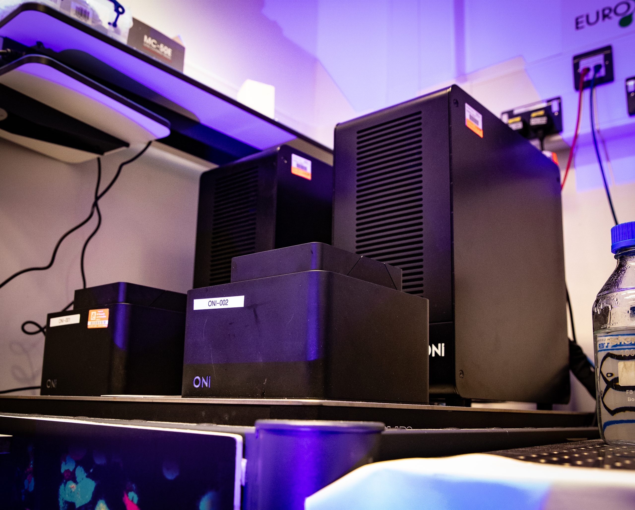
At Octopus we have three super-resolution Nanoimagers (ONI). All three are available for the Octopus-developed FLImP technique, while one can be used for extracellular vesicle and liquid nanoparticle characterisation using dSTORM.
The Nanoimager supports multiple imaging methods, including dSTORM, PALM, DNA PAINT, single particle tracking and single molecule FRET. Its adjustable illumination angles (0 to 65°) allow for epifluorescence, HILO, and total internal reflection fluorescence (TIRF). In TIRF mode only fluorophores within approximately 200nm of the coverslip are excited, which greatly reduces background and allowing surface specific imaging. Epifluorescence (widefield) microscopy uses a parallel beam of light to illuminate the entire sample, making it well suited for thicker specimens (>10 microns). However, this broad illumination also excites molecules outside the focal plane, increasing background. In contrast, TIRF selectively excites fluorophores near the coverslip surface, reducing background noise. This makes TIRF especially powerful for studying fine structures close to the coverslip, as well as molecules attached to membranes or surfaces.
High-power lasers allow simultaneous two-colour imaging, with sample power densities up to 4kWcm⁻². Single-molecule localization microscopy achieves ~20nm lateral and ~100nm axial resolution. For live-cell imaging, the system provides precise temperature control up to 42°C with ±0.1°C stability.