MINFLUX microscopy

Ultra-high precision 3D localisation microscopy and 2D single molecule tracking.

The instrument employing this technique is the Abberior Instruments MINFLUX.

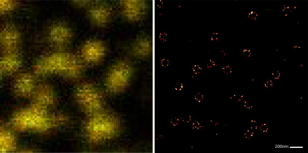
Overview of the technique

MINFLUX is a new super-resolution fluorescence imaging technique that can measure the positions of single fluorescent dyes with outstanding precision. Samples labelled with stochastic blinking dyes are probed with a doughnut shaped excitation beam in a rapidly shrinking search pattern that locates the dye position. This allows the position to be determined with a precision of approximately 2nm (2D measurement) or 3 to 4nm (3D measurement) using relatively few fluorescence photons from the limited photon budget of the dye. Dye blinking can be achieved using photo-switching cyanine dyes, photoactivatable dyes or the DNA-PAINT method.

MINFLUX can also track moving dyes in 2D with unprecedented localisation precision and temporal resolution. For example, our MINFLUX system has tracked fluorescently tagged proteins with 4nm average localisation precision and 1.6ms average time jumps. Time jumps of 120 microns with 20nm localisation precision have been reported tracking fluorescently tagged lipids (Schmidt et al. 2021), as has the stepping motion of kinesin (Deguchi et al. 2023), dynein (Schleske et al. 2024).

Comparative images of Nup96 complexes labelled with Alexa 647. Left: standard confocal mode. Right: 2D Minflux mode.

Applications

The applications of MINFLUX microscopy include:

  • intracellular structures: nucleopore, centrioles, microtubules
  • receptor protein oligomer structure
  • motor protein dynamics
  • membrane protein diffusion

MINFLUX imaging is highly suited to structural determination of recurring stable cellular structures and macromolecular complexes such as nucleopores, centrioles and microtubules. Observation of receptor protein oligomers is possible in theory but made considerably harder in practice by the limited efficiency of fluorescent labelling technologies. MINFLUX works well with in vitro samples or adherent cells prepared on glass coverslips, eight-well chamber slides and 35mm glass bottomed dishes.

Using our MINFLUX we have also successfully tracked proteins diffusing on the surface of live CHO cells and bacteria, as well as motor proteins moving along microtubules.