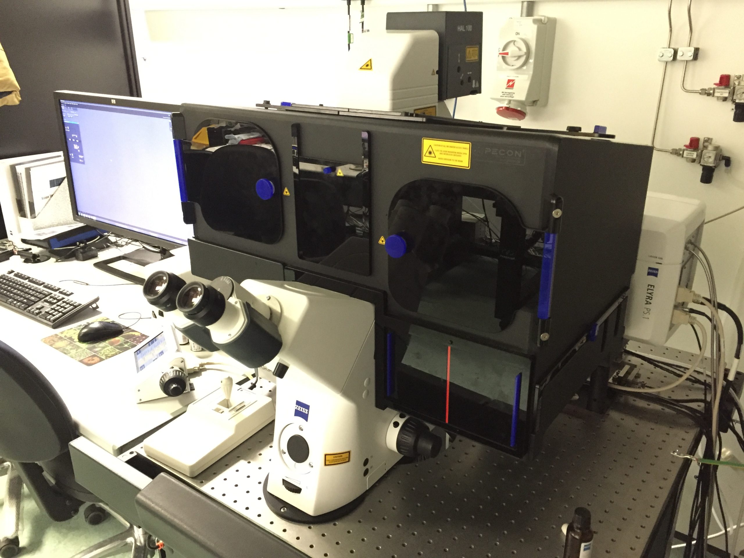
Overview of the technique
Structured illumination microscopy (SIM) is a method of imaging fluorescently labelled structures at greater spatial resolution than a conventional fluorescence microscope is capable of optical diffraction imposes a limit of approximately 200nm lateral and 500nm axial resolution in the conventional microscope, both of which are doubled in SIM. It involves imaging a grating onto the sample to produce Moiré fringes, which may be resolved in the microscope even if the sample structure cannot be seen directly.
As with other super-resolution techniques, such as single molecule localisation microscopy (SMLM) and stimulated emission depletion microscopy (STED), structured illumination microscopy (SIM) enables fine structure to be resolved. The advantage of SIM over other super-resolution imaging techniques is that the choice of fluorescent dye is limited solely by the laser lines available to excite it. It also uses much lower laser powers and hence is suited to applications where limiting phototoxicity is important or for time-lapse imaging. It is compatible with multicolour imaging of up to four fluorescent probes. SIM can be performed on the same microscope as SMLM, enabling correlative imaging with the two super-resolution methods.
SIM is well-suited to imaging live cells because it has low phototoxicity and the imaging duration is low, of the order of seconds. It is equally applicable to animal and plant biology, as well as in chemistry and materials science.