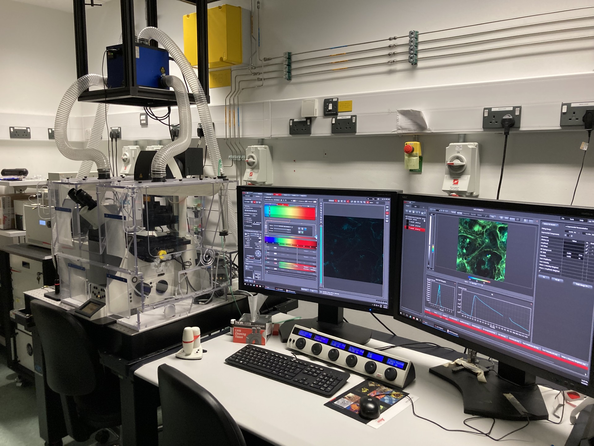
Overview of the technique
Stimulated Emission Depletion (STED) microscopy is a super-resolution imaging technique that overcomes the diffraction limit of light, allowing for imaging at resolutions beyond conventional optical microscopy. In STED, a focused laser beam is used to excite fluorescent molecules in the sample. A second, “depletion” laser with a doughnut-shaped profile then selectively depletes fluorescence in the outer regions of the excitation area, effectively limiting the fluorescence emission to a small central spot. This spatially confined emission enables the imaging of structures at resolutions down to the nanometre scale, far surpassing the traditional diffraction limit of approximately 200nm.
By carefully controlling the timing and intensity of the depletion laser, STED microscopy enables imaging of dynamic biological processes with exceptional spatial precision. It has become a powerful tool in fields such as cell biology, neuroscience, and materials science, where studying fine details like protein interactions, cellular structures, and molecular dynamics at nanometre resolution is critical. Despite its complexity, STED is gaining popularity due to its ability to provide high-resolution imaging without the need for complex sample preparation or electron microscopy.