Overview of the technique
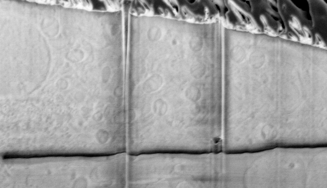
FIB-SEM vEM, also known as slice-and-view imaging, is an imaging technique that can obtain volume data from room temperature or cryogenic samples over tens of microns with voxel sizes down to the nanometre scale.
This technique works by opening a trench in the sample to create a trench face, which is then imaged in cycles by removing material (milling) an nm-thick slice of a sample using a focused gallium ion beam and then imaging that same area using a scanning electron microscope. The process is repeated cycle after cycle, destroying that area of the sample in the process. This technique is one of many volume electron microscopy (vEM) techniques and is regularly used to image biological samples, biocomposite materials and even metals or ceramics.
Compared to other vEM techniques, FIB-SEM vEM allows the imaging of a smaller area, but at higher resolution, and often with isotropic voxels, which is a boon for analysis.
At CLF Octopus we can perform FIB-SEM vEM of samples at room temperature (typically blocks of biological samples stained with heavy metals and embedded in resin) or at cryogenic temperature (typically samples plunge-frozen on EM grids or high-pressure frozen in planchettes or on sapphire disks).
Applications
Some applications of vEM include:
- viral infection studies – depending on the sample, viral particles can be imaged and counted individually
- biological effects of particulate material in tissue and culture
- organelle ultrastructure in health and disease
- biomineralisation of marine organisms
- visualisation of organelle interactions in signalling
- bacterial interactions with nanomachined or functionalised surfaces
Samples and general information
Sample sizes can be very large (as long as they fit in the chamber; see specifications), but they must have a flat accessible surface for milling and imaging. Contrast is provided by heavy metal impregnation for room temperature samples and by charging differences between aqueous and lipidic structures for cryo samples.
Of note, cryogenic samples provide much less contrast and are not a suitable first approach to vEM for most beginners, but they provide ultrastructural information without the need for chemical fixation. However, samples containing CL2 and above viruses or other pathogens must be fixed before cryogenic preparation and data proving that the treatment abolishes infectivity must be provided.
Typical imaged volumes are 40 x 30 x 30 micron at 10 x 10 x 10nm voxel size, but larger volumes can be imaged, and smaller voxel sizes can be obtained, depending on the sample. This technique typically requires 24 to 48 hours per sample for cryogenic experiments and up to an entire week per sample for RT samples, depending on imaged volume and voxel size requirements. This technique is not generally suitable for sample screening.
Support
We provide our users:
- support and training for sample preparation for cryo-vEM (plunge-freezing on EM grids)
- support and training for room-temperature single-modality or correlative imaging of biological and material specimens, 2D or 3D
- support and training for cryogenic temperature single-modality or correlative imaging of biological and material specimens, 2D or 3D
- secure data storage and transfer
- support and training for data analysis using open-source environments such as Fiji, Icy and the MIB-3D Slicer-Blender pipeline
We do not have facilities for metal impregnation and resin embedding of biological samples. Samples for imaging at CLF of this nature must be prepared offsite in advance. If you do not have the required facilities in house, please let us know as we may be able to make introductions to collaborators who may be able to help.
Essential reading
An essential resource for vEM is the VolumeEM website, a community platform that collects all the information about vEM, including FIB-SEM slice and view. Good starting points for your experimental planning from this website include: