Using ultrafast imaging; Jonathan Wood and a group of scientists at Gemini captured images of laser driven shock waves in a silicon target, enabling bright X-ray probing of high energy density physics without the use of larger, light synchrotron sources.
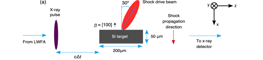
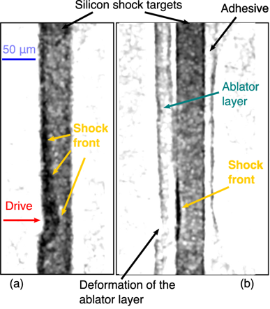
High energy density physics (HEDP) experiments constitute research in fields ranging from condensed matter to meteorite impacts, from material failure mechanisms to the behaviour of planetary cores. States such as these, with high temperatures and pressures, are accessible in the laboratory through dynamic compression experiments – a shock wave is driven into a material, with this particular experiment, by a pulsed laser.
For more than a decade at Imperial College London, Dr Stuart Mangles and Dr Daniel Eakins (who proposed the research), along with a research team have been developing a new type of compact accelerator called a laser wakefield accelerator which has been used in this experiment. It has the capability to make ultrafast flashes of X-rays that can ‘freeze’ the motion of rapidly changing systems.
“The X-rays produced by the laser wakefield accelerator at Gemini are great for taking snapshots of shocks as they fly through some material because they are very short flashes of X-rays.”
Dr Stuart Mangles (Imperial College London)
Furthermore, the use of betatron radiation – ultrashort pulsed, hard, bright X-rays – for radiography is an enticing foray into the imaging of rapidly evolving phenomena. As it stands presently, traditional surface based measurements are proving inadequate as the subsurface interactions cannot be diagnosed directly. The result being, measurements of shock properties at the surface are potentially affected by interactions below the surface which are not being observed.
The Gemini laser at the Central Laser Facility, with its two beamlines, proved invaluable as the Gemini South beam was used in the production of the betatron radiation and the Gemini North beam was used to produce the shock wave in the target. The delay between the target being hit and the betatron probe was able to be varied continuously between 0 and 12 nanoseconds allowing for snapshots to be taken of the shock wave. One benefit proposed by betatron radiation is that rapidly evolving phenomenon can be imaged without the need for larger, more expensive, light synchrotron sources.
The footprint of the experiment detailed, had the target placed 0.12m from the source and the camera was 3.70m from the source. When considering the scale of X-ray imaging beamlines from synchrotrons, which are typically around 100m long, the benefit of using an experimental setup this small is immense. It allows ultrafast, hard X-ray probing experiments to be performed at university scale facilities and at a fraction of the cost! Alternatively, a betatron imaging beamline could be implemented at large scale HEDP facilities, again lowering the cost. Further still, due to the high flux of betatron X-rays, standard detectors were sufficient to allow single pulse imaging.