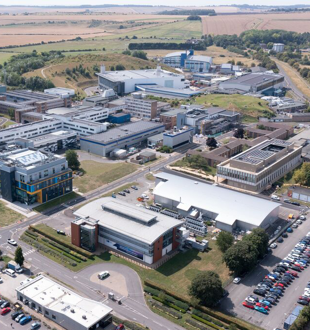
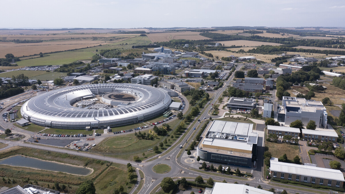
Developing technologies
Our aim is to provide new techniques for the user community that help to link molecular structure and function in the environment of the cell.
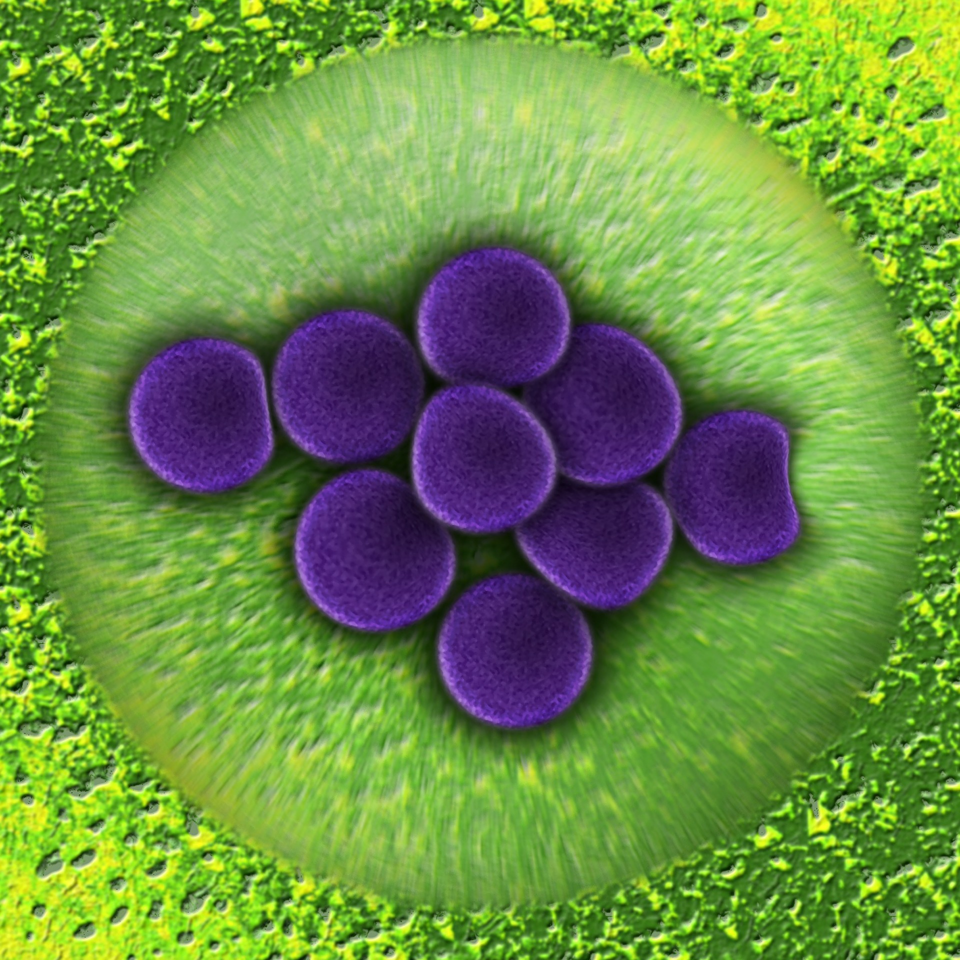
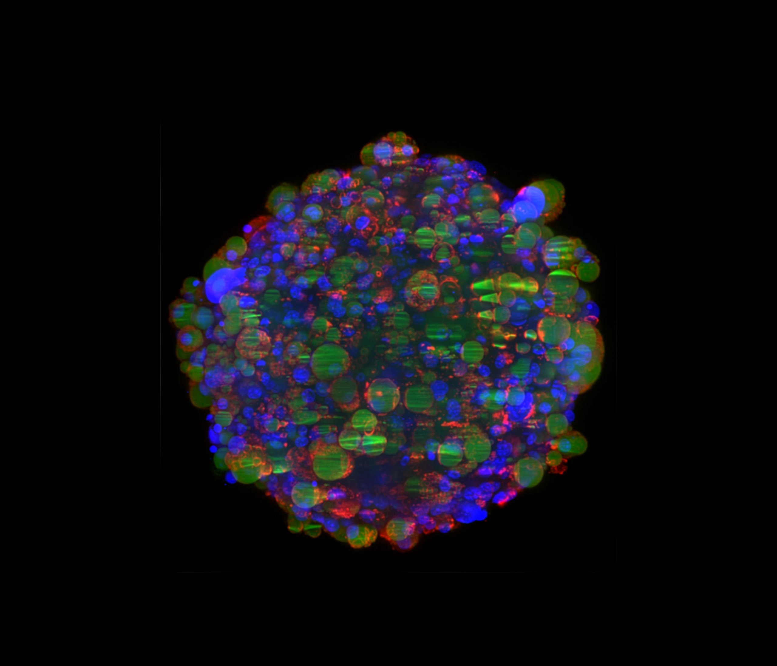
Fluorophore Localisation Imaging with Photobleaching (FLImP)
FLImP is an automated single molecule imaging technique capable of resolving separations between fluorescently tagged molecules as small as 5nm and with about 5 nm precision.
This technique is particularly powerful for resolving the architecture of molecular complexes in cells, and has resulted in two publications in Nature Communications that have helped to overturn current understanding of activation of the Epidermal Growth Factor Receptor.
FLImP is now being applied by user groups working in other areas (galectin-3-fibrosome and endocytosis).
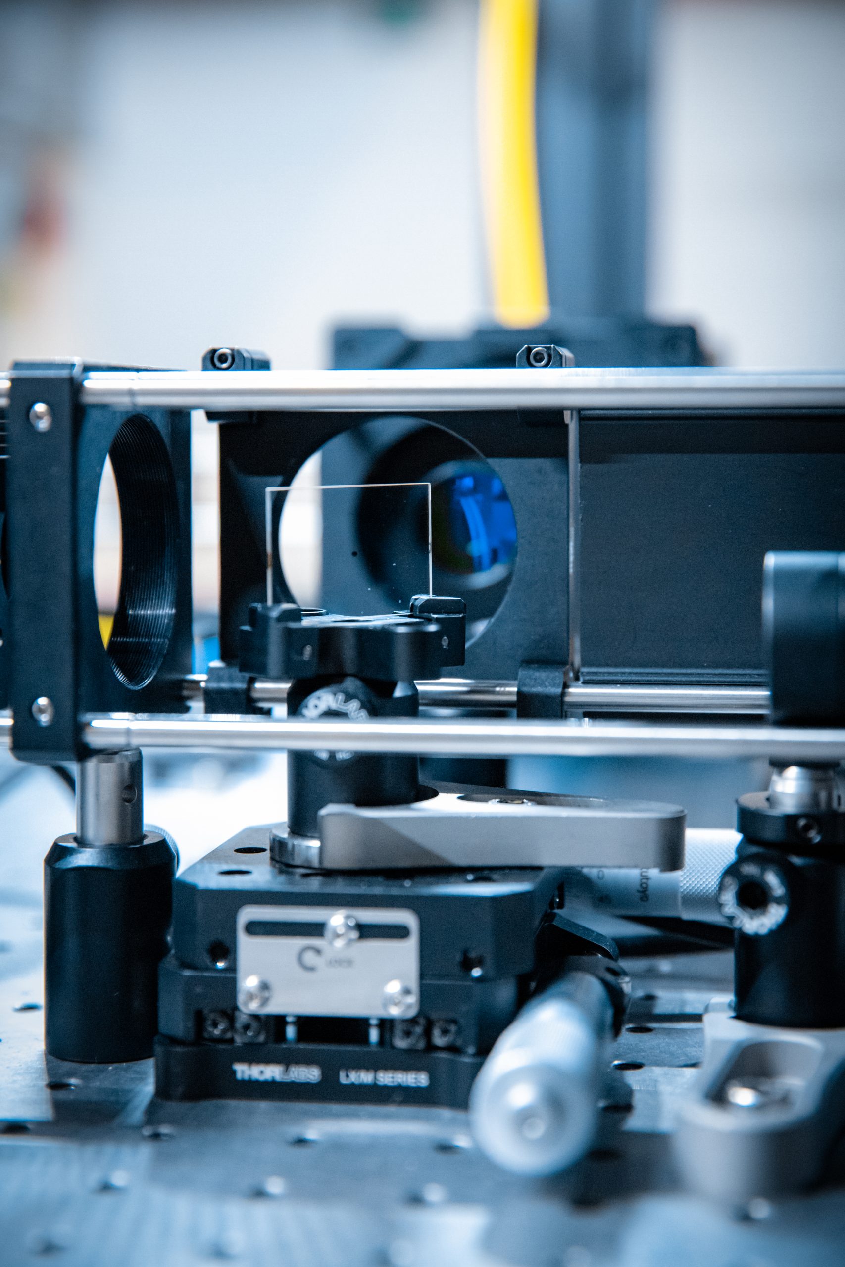
Cryogenic Stochastic Optical Reconstruction Microscopy (Cryo-STORM)
A major area of technology development in biological microscopy is super-resolution microscopy at cryogenic temperatures.
The objective of this development is:
- to improve both resolution and sample preservation
- to pave the way towards more effective correlative light and electron microscopy
Super-resolution microscopy at cryogenic temperatures is challenging because of the need for a high numerical aperture objective lens, usually achieved by the use of oil immersion lenses that do not work at low temperature, and the requirement to control chemically the photo-physics of the fluorescent labels using special buffers, again impossible with frozen samples.
The CLF has developed cryo-STORM microscopy using aplanatic solid immersion lenses (ASILs) to achieve high NAs at cryo temperatures, and special illumination and data acquisition techniques to allow single molecule localisation in the absence of special buffers.
Cryo-STORM has now been accessed by several external groups investigating receptors, membranes, and new fluorescent probes.
Plasma-Focused Ion Beam (pFIB)
As part of the correlative microscopy development programme, a cryo plasma focused ion beam scanning electron microscope (pFIB) has been purchased. This will be used mainly for the production of lamellas for TEM imaging in collaboration with eBIC at Diamond Light Source.
It is also intended to extend imaging at cryogenic temperatures to MINFLUX, bringing the resolution of optical microscopy closer to that of the electron microscope. Correlation of structural data from TEM with nanometre-resolution optical imaging is a step towards enabling in situ structural biology and will attract a new user community to the CLF.
Software development
These biological microscopy developments above have been enabled by a significant effort in data acquisition and analysis, including the use of machine learning and artificial intelligence.
The efficiency of the FLImP method has been greatly increased by the automation of both data acquisition and data analysis, with a greater than 10-fold improvement in time from sample loading to obtaining useful data.
The analysis is computationally intense and requires the use of cloud computing services in STFC and elsewhere.
Co-location of the CLF with STFC Scientific Computing infrastructure and expertise has been useful for these developments.
For example, the CLF has a strong collaboration with STFC’s Scientific Machine Learning Group. This has led to the development of automated methods for segmentation of image volumes from 3D FIB-SEM and will contribute to the CLF’s development of new CLEM methods and workflows.
The Scientific Machine Learning Group and the wider Scientific Computing are heavily involved in the design of the high repetition rate data management system, as well as in developing customised solutions for data analysis tools, like real-time Computed Tomography, for the EPAC facility.