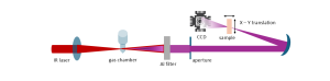
XUV ptychography is an innovative imaging technique that enables researchers to visualise structures at the nanoscale with exceptional resolution. Utilising XUV light, which is characterised by its very short wavelength, this method enables higher resolution than conventional optical microscopy.
In practice, a coherent XUV beam is scanned across the sample while a detector records a sequence of overlapping diffraction patterns. Advanced phase retrieval algorithms then reconstruct a high-resolution image from the dataset, revealing the morphology and, in some cases, the composition of the specimen. Both amplitude and phase information are retrieved.
The method is proving valuable across many research domains. In condensed matter physics, it can reveal nanoscale ordering and defects in quantum materials. In chemistry, it enables the study of catalytic surfaces and molecular assemblies. In the life sciences, it can resolve subcellular structures while minimising radiation-induced damage. Its non-destructive nature, high sensitivity, and capacity for quantitative analysis make it especially well-suited to delicate or complex specimens.