Leica TCS SP8 X Digital Light Sheet (DLS) Multiphoton (MP)

The techniques employed on this instrument are

Experiment information – Light sheet mode

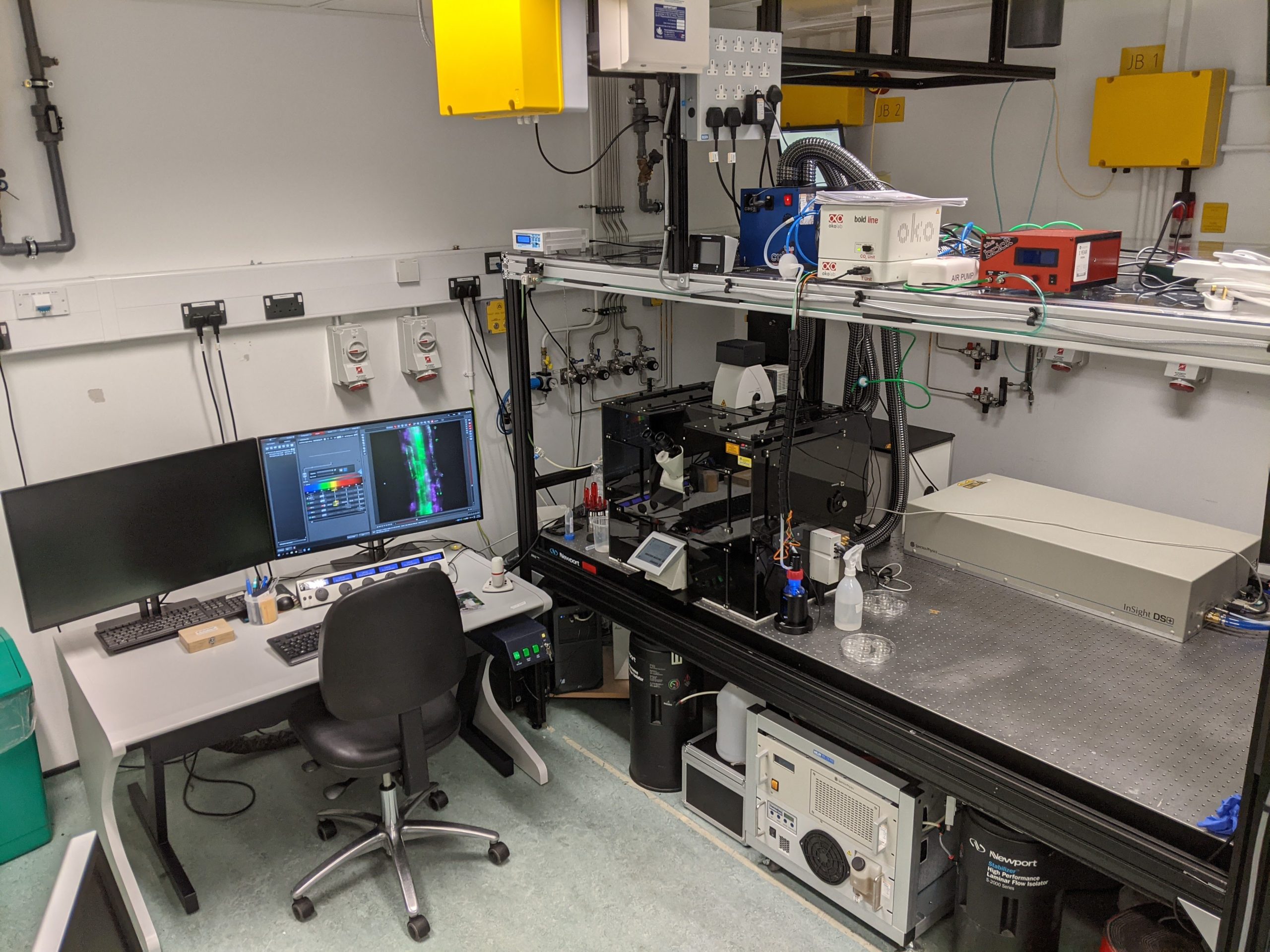
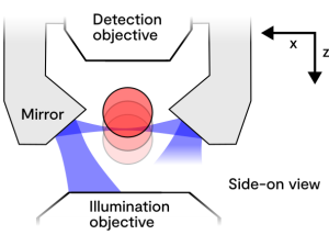
The Leica SP8 is capable of light sheet imaging of live, fixed or cleared samples. For live samples, there is an Okolab environmental chamber for humidity, CO2 and temperature control. The sample size is limited by the TwinFlect mirrors which come down either side of the sample to project the light sheet across (see figure below), and the sterility of the sample environment is compromised by the mirrors, which should be factored in when planning long-term experiments.

Sphero​ids and organoids are perfect for this system, along with small transparent organisms (for example, larval zebrafish) where only a small area (<1mm) needs to be imaged. Additionally, cells growing inside of a 3D gel are suitable for this system. We have also used it for more innovative applications such as imaging cells flowing through tubing or nozzles during bioprinting.

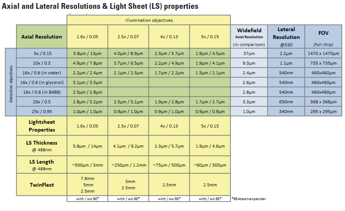
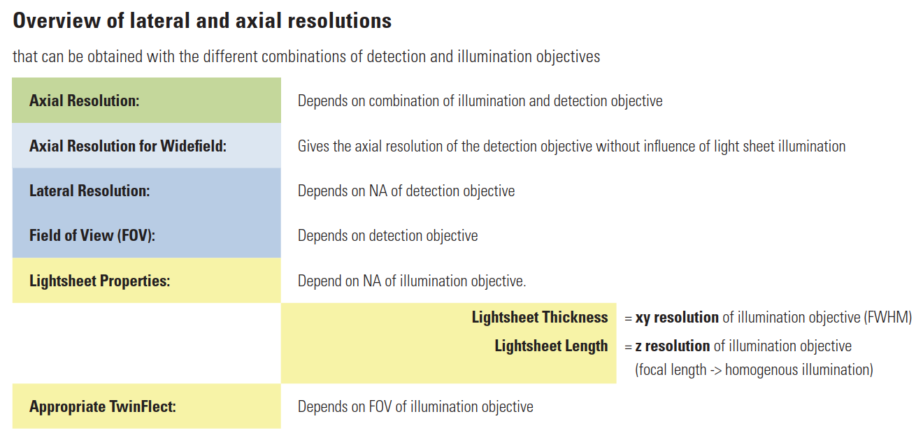
There are a variety of configurations listed below, but we generally use the 1.6x or 2.5x illumination objective coupled with the 10x or 16x detection objective to give the best compromise of resolution with field of view (see graph in the specifications below). Discussion with a link scientist will help to determine the best configuration for your samples.

Experiment information – Multiphoton and confocal modes

The Leica SP8 is capable of confocal and multiphoton imaging in an inverted setup (the objectives are below the sample) which is most well-suited to slides or coverslips and dishes with glass bases. Typically, the confocal and multiphoton modalities are used as an accessory to other techniques in the facility. There is a heated chamber for environmental control of CO2 and temperature. We have carried out second harmonic generation (SHG) imaging of collagen in skin models, mammalian intervertebral discs and biomaterials, as well as multiphoton imaging of skin and chemical formulations.

There are a variety of laser lines, objectives and detectors available (see technical specifications below). Discussion with a link scientist will help to determine the best configuration for your samples.

Technical specifications

  Light Sheet  Multiphoton/Confocal 
Objectives  Detection:5x/0.15 IMM, 10x/0.3 W, 16x/0.6 IMM, 25x/0.95 W 

Illumination: 1.6x/0.05, 2.5x/0.07, 5x/0.15 

Confocal:10x/0.4, 20x/0.75 IMM, 40x/1.1 W, 63x/1.2 W, 100x/1.4 Oil 

Preferred multiphoton:25x/0.95 W 

Lasers  NKT SuperK Xtreme WLL (470-670 nm), Diode 405 nm  NKT SuperK Xtreme WLL (470-670 nm), Diode 405 nm, OPO InSight DeepSee (680-1300 nm) 
Filters  455-495, 504-545, 575-615, 575-635, 510-536+597-623 (reduce chlorophyll autofluorescence), 660-734,  500-632+650-750, and a quad band with notches for 405/488/561/633nm  Internal detectors:Free-choice 380 to 800nm 

External detectors (multiphoton): 435-450, 435-485, 467-499, 500-550, 565-605nm 

Detectors  Hamamatsu ORCA Flash 4.0 V2 sCMOS camera up to 30fps full-frame, faster if cropping. For resolution, see below table.  External detectors: 2 PMT, 2 HyD 

Internal detectors: 2 PMT, 2 HyD 

External transmitted detector 

Field of view  Up to 1mm  Up to 1mm 
Sample size and mounting  Up to 4mm wide, 1.5mm thick, 20mm long. Usually mounted to glass-bottomed dishes by glue, capillary tube, U-shaped glass capillary or inside agarose wells. Alternatively, there is a larger mounting frame or rotating mounting frame to rotate capillaries.  Sample size is not limited, coverslip or glass-bottomed dish for flat-mounting for inverted objectives 
Refractive index 

compatibility 

(cleared samples) 

1.33-1.57 for the 5x and 16x objectives   Not suitable for cleared samples 
Temperature  From room temperature to 60°C  From room temperature to 37°C