Light sheet microscopy

Capturing fast dynamics gently or structure across large volumes. The instruments employing this technique are:

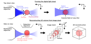
Overview of the technique

Light sheet microscopy is a fluorescence imaging technique with a growing number of specialised variants, each tailored to specific applications. At Octopus, we use digitally scanned laser light sheets. In this method, a Gaussian laser beam is directed at the sample from a direction perpendicular to the detection axis. The beam is rapidly scanned along the y-axis, much faster than the camera’s exposure time, creating a full plane of illumination that is captured in a single image. By moving the sample through the light sheet, a stack of images can be acquired plane by plane, allowing for 3D reconstruction of the fluorescence signal.

Selected publications

See the following papers for more information about light sheet microscopy:

Light sheet fluorescence microscopy


Light-sheet microscopy: a tutorial


Tutorial: practical considerations for tissue clearing and imaging

Relevant contacts

Robert Lees

Link Scientist

Robert provides scientific and technical expertise and support to academic and industrial partners for all aspects of their microscopy experiments, from design to acquisition and analysis.

Sarah Needham

Senior Link Scientist

Sarah joined STFC as a postdoctoral researcher in 2005 and the CLF in 2008.